
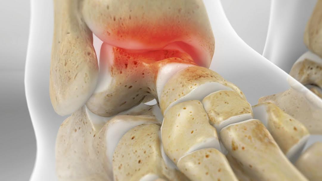
ANKLE CARTILAGE INJURY
The ankle joint is the articulation formed between the end of the tibia and fibula (shinbones) with the talus (ankle bone). Osteochondral injuries are injuries at the joint level, characterized by damage to the cartilage as well as bone. The top part of the talus and/or the lower end of the tibia can be affected.
Osteochondral injuries are most often caused by trauma to the ankle joint, such as ankle sprains. Some cases may not have any previous history of ankle injury and may be caused by local osteonecrosis or a metabolic defect.
Symptoms can be:
-
Localized pain of the ankle joint
-
Tenderness and swelling of the ankle joint
-
Difficulty in weight bearing
-
Locking of the ankle
Osteochondral injuries are diagnosed by physical examination, X-ray and CT and MRI scans. X-ray images can reveal other fractures, bone spurs and narrowing of the joint. A CT scan helps to identify any loose bony fragments in the joint and cysts located at the end of the bones. MRI is the best imaging modality, which helps to visualize the cartilage and bone lesions as well as bone oedema.
Regarding therapeutic options, non-surgical or surgical treatment may be recommended for the management of osteochondral injuries of the ankle joint. Non-surgical treatment with immobilization, restricted weight-bearing and physical therapy may be ordered. This will help the bone and cartilage to heal and improve muscle strength, mobility and coordination.
Surgical treatment is recommended for more severe injuries and those which fail conservative treatment. Ankle arthroscopy will be performed and the surgical options are:
-
Debridement (removal) of the damaged cartilage and removal of any loose bodies
-
Microfracture (small perforations in the bony defect) of the lesion
-
Grafting of cartilage and bone
-
Fixation of the fragments (when large enough) with the help of screws