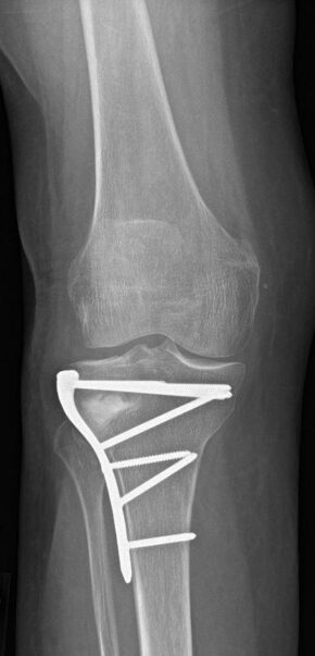
During the surgery, an incision or cut is commonly made on the inner aspect of the knee. Before the surgery, calculations are made to plan how much change in alignment of the leg is needed. A cut is made into the tibia bone and then opened, to make the change in alignment. A bone wedge is inserted between the bone fragments, which helps the bone to heal across this bone wedge. A plate is used to hold the knee bone alignment and then fixed with screws.
Post-operative care
After osteotomy surgery patients may require one to two days of hospitalisation. Immediately after surgery, a light compressive knee brace or splint may be used to protect the knee and pain medications are given to reduce pain or swelling. During this time patients are instructed to follow post-operative protocols for a successful outcome.
Post-operative instructions include the use of TED stockings to reduce the risk of clot formation, crutch walking for at least 9-12 weeks and physiotherapy. Physiotherapy includes range of motion and strengthening exercises, to restore range of motion and improve muscle strength. Physiotherapy may begin the day after surgery to help the patient to move in and out of bed and to learn to use crutches in a safe manner. Crutch walking involves using crutches without weight bearing on the operated leg immediately after the surgery to allow the bone to heal. After a few weeks, progression to partial weight bearing in the operated leg will be encouraged.
Complications
As with any surgery, patients undergoing this surgery are at risk for developing certain complications following the surgery. Common risks and complications of the surgery are pain, swelling, numbness or injury to the surrounding nerves or vessels, infection, compartment syndrome, deep vein thrombosis, loosening of instrumentation (plates and screws) and non-union of the bone. Subsequently, a knee replacement may still be required.

